Not sure if this plays but it's cute. Myrna rolls over and "plays cute."

Our cat Myrna Loy, born March 22, 2009, died of HCM 8/19/15. We will continue the blog and FB page. With five cats, we often have a variety of medical issues. Please share your information. Please check the Archives, Categories, Search, and Tabs for more information.
Wednesday, July 27, 2011

Busy Week This Week with Vet Visits
Just an update on our cats:
Myrna Loy is doing great. Bounced back from two weeks ago CHF; taking Buprenex as a pain med. Keeping her calm and cool.
Elizabeth Taylor keeps getting hit with idiopathic cystitis. We had her on Gabapentin last week because we were running out of Buprenex and were in the middle of getting it compounded into a transdermal version. I was saving the oral Buprenex for Myrna. The first two days Elizabeth was on Gab, she was fine. I administered .40 ml. She slept but wasn't out cold as she was two weeks ago when I gave what was prescribed-.60. On .40 she ate well; responded to touch, was alert, and did sleep some. But by the third day, she seemed cranky and in pain. By the fourth day, she was just not happy. When she came off the med, she seemed to crash-not sleeping crash, but I'm angry and not feeling well crash. By the fifth day-and I had to wean her off so by the third night I reduced her med-she just seemed miserable. Her check-up was yesterday. She was in such a foul mood that she hissed, growled vehemently, swatted, and then finally bit me on the arm while I held her or moved her from the carrier!! That is just not my Lizzie! She's a sweet girl. She never bites me for any reason. Last night, we did not give her a dose of Gab and today she's doing so much better!! She's calm, she's her old gentle self. We will start her on the transdermal Buprenex on Thursday or Friday. Gabapentin is a seizure med and it might work well for those patients, but doctors also use it for neuro pain. I don't know what Gab is suppose to do but it's not good for solving pain. The side effects are too costly.
Cooper's hypercalcemia is coming down slowly with fiber, magnesium and potassium supplements. We also give extra water and extra protein source (all from various human websites that say this might help.)
We can't tell what level of Prozac works for Jimmy. I had him at .110 and then .109 to see how low he could go. But we still had out of the box issues-but not all attributed to him. What we do know is that we haven't seen him go or evidence of him going. So, this week he's up to .112. He seems much more relaxed and engaged and playful. But still, we can't see evidence of him using a box. But I can't find any out of the box issues, also. Just where is his urine?
Myrna Loy is doing great. Bounced back from two weeks ago CHF; taking Buprenex as a pain med. Keeping her calm and cool.
Elizabeth Taylor keeps getting hit with idiopathic cystitis. We had her on Gabapentin last week because we were running out of Buprenex and were in the middle of getting it compounded into a transdermal version. I was saving the oral Buprenex for Myrna. The first two days Elizabeth was on Gab, she was fine. I administered .40 ml. She slept but wasn't out cold as she was two weeks ago when I gave what was prescribed-.60. On .40 she ate well; responded to touch, was alert, and did sleep some. But by the third day, she seemed cranky and in pain. By the fourth day, she was just not happy. When she came off the med, she seemed to crash-not sleeping crash, but I'm angry and not feeling well crash. By the fifth day-and I had to wean her off so by the third night I reduced her med-she just seemed miserable. Her check-up was yesterday. She was in such a foul mood that she hissed, growled vehemently, swatted, and then finally bit me on the arm while I held her or moved her from the carrier!! That is just not my Lizzie! She's a sweet girl. She never bites me for any reason. Last night, we did not give her a dose of Gab and today she's doing so much better!! She's calm, she's her old gentle self. We will start her on the transdermal Buprenex on Thursday or Friday. Gabapentin is a seizure med and it might work well for those patients, but doctors also use it for neuro pain. I don't know what Gab is suppose to do but it's not good for solving pain. The side effects are too costly.
Cooper's hypercalcemia is coming down slowly with fiber, magnesium and potassium supplements. We also give extra water and extra protein source (all from various human websites that say this might help.)
 |
| Jimmy after playing in the closet on the rolled up rug. |
Poducts and Pictures from St. Louis Vet Conference
 |
| Vendor hall |
 |
| Inside vendor hall St. Louis vet conference |
 |
| Freebies! Left-plaque remover for pets; urine-away by Felawy; and Senior cat litter by makers of Prescious Cat litter-crystals that don't stick to cats. |
 |
| Gets rid of plaque on pets' teeth. Good option for pets who cannot get anesthsia for dental cleaning. |
 |
| New harness/leash set-hooks around chest not neck. More comfortable. |
 |
| Doggie was free; clicker is for cat training; blue disc is a treat toy-put in treats; cats are suppose to play w/it and get out treats. |
 |
| New sand like urine sample collection process-put sand in box; cat goes on it; urine stands on sand instead of sinks in. |
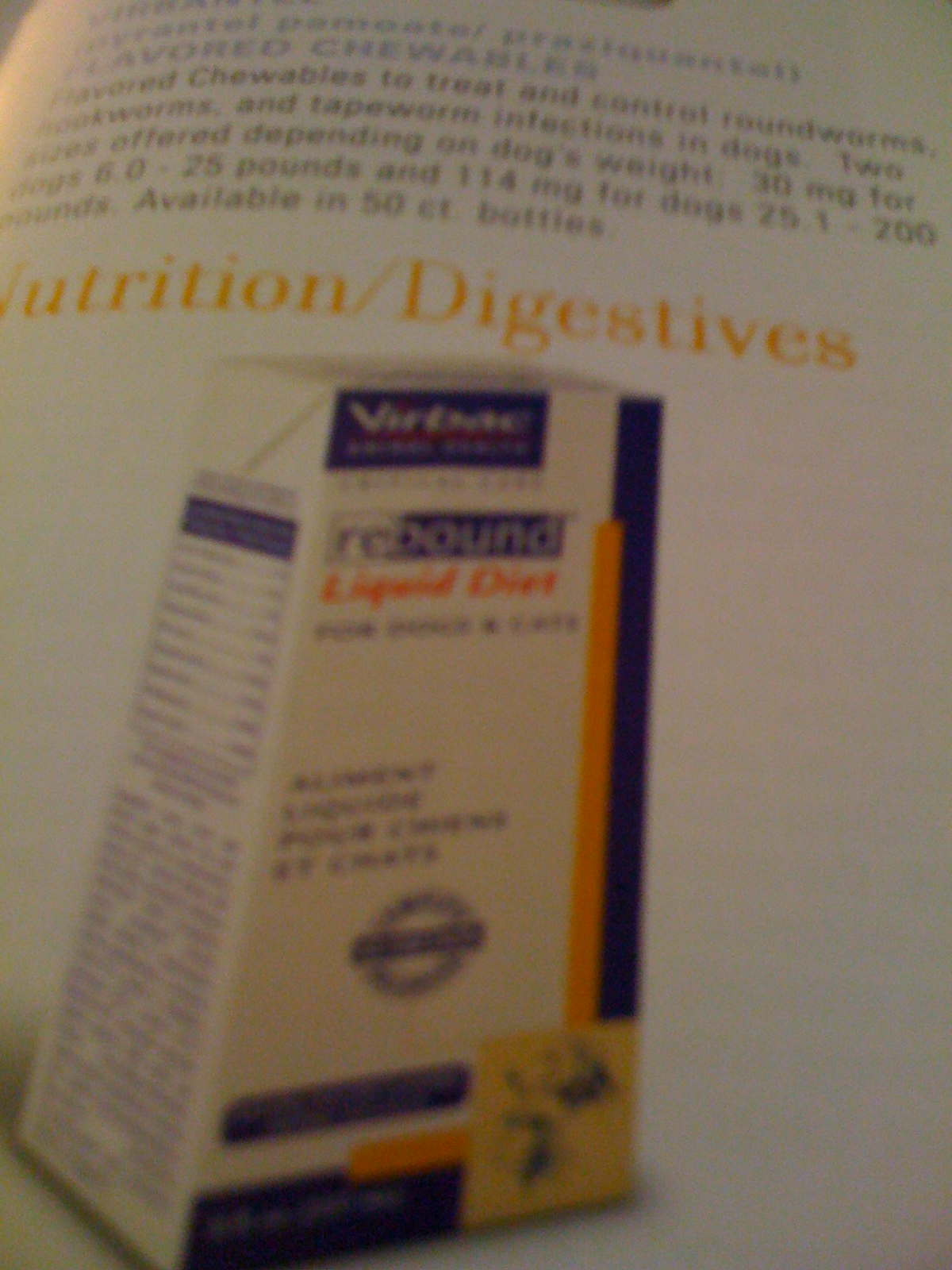 |
| Good source of nutrition for cats who are ill, recovering, etc. Might also be good for feeding tubes. |
| Add caption |
 |
| Felaway has a "Dear Tabby" Facebook page |
 |
| New procedure for sub-q fluids-put stent under skin along spine (no pain to pets) to give fluids. Good for those pets that need daily sub q. Makes it easier to do. |
 |
| New urine tester in petrie dish. Colors tell you what infection urine has. |
 |
| Fish oil refined. Doesn't smell like fish. Tastes better than fish oil capsules. |
 |
| For dogs-socks. They're so cute! For all sizes. Wicks away moisture. Helps dogs to get traction on slippery floors. Protects from hot asphalt and concrete sidewalks. |
Sunday, July 24, 2011
Notes from July 2011 St. Louis AVMA Conference-HCM
These are papers from the July 2011 St. Louis AVMA conference. I’ve edited them to delete some of the vet only jargon and for space considerations. If you see (…) that means that information has been deleted due to these considerations. The ones listed here include HCM, and other heart related papers, treatments, papers on x-rays and echos, thrombosis, kidney disease, idiopathic cystitis, pain management, anesthesia and cardiac disease, supplements and other hazards for pets, and some other basic information I hope is helpful.
My note-regarding the paper below, in class she said that there is a wide range and a non-cohesive approach to treating HCM among vets. There isn’t much data available on treatments and what slows the progression of the disease. Many vets follow human studies to gain more knowledge of the disease. There is research on a drug in this or that study but not regarding one total approach. Part of the problem (aside from financing a study) is that each pet presents with different side effects of the disease which requires an individual approach to the issue. But due to a lack of research, many doctors hesitate to use multiple drugs and multiple approaches. 8% or less of cats have HCM. More males than females will get the disease. There are breeds that will get it more, such as Maine Coon and Ragdoll. The average age of detection is 6-16 years. Half of the cats have no symptoms but are diagnosed due to other unrelated issues-before a dental or surgical procedure for example. Or others become sick from medications given for other illnesses, such as steroids, and CHF kicks in. Usually lethargy and difficult breathing is what owners notice in the cats. Up to 34% of cats have heart murmurs but not HCM. Most cats that will get HCM are born with a gene that will cause HCM. Others will get it due to having another disease which brings on the condition or brings on CHF. Sometimes the onset of the disease is greatly delayed for unknown reasons. But if the cat has the gene, the disease will eventually form. A good and inexpensive diagnostic tool is the x-ray. If an owner cannot yet afford an echo, an x-ray will show if the heart is enlarged. If the heart is enlarged, then an echo is needed. But if the heart is not enlarged and the cat does not present with other symptoms, then carefully monitor in the future with more x-rays until an echo is warranted. She discussed in class (but not in the paper below) how to measure the heart via x-ray. It’s too complicated for me to explain but has something to do with measuring the size of the heart in relation to the vertebrae by taking different mathematical measurements at different axis points on the x-ray to determine how much of the chest cavity the heart is taking up. If it is up to 8 or less vertebrae there is not an enlarged heart yet. If it is 9 or more, than an echo is warranted, the heart is enlarged, and treatment must begin. More doctors need to be aware of this as do owners of pets. However, having an HCM cat, I understand the necessity of getting an echo and will always recommend one for those cats who are presenting with HCM related side effects (such as lethargy, breathing difficulty, etc.) The only study she cited that showed which drug actually worked for HCM was the study regarding lasik. All other drugs have had either no feline studies or have not shown to work for all cats. But she said that certain things must be treated-tachycardia, left atrial enlargement, and severe dynamic stenosis. She gives ace inhibitor such as enalapril. Beta blockers such as atelenol in human studies have proven effective and she gives it to cats to treat tachycardia. She agreed (in a Q&A) with an audience member that vitamins were needed.
There is a school doing a genetic study in cats. If you want to find out more, call 909-513-8279 or go to www.cvm.ncsu.edu/vhc/csds/vcgl/index.html
Feline Hypertrophic Cardiomyopathy Update
Meg M Sleeper VMD, DACVIM (Cardiology)
Associate Professor of Cardiology; University of Pennsylvania School of Veterinary Medicine
Hypertrophic cardiomyopathy (HCM) is a primary myocardial disease that affects left
ventricular (LV) myocardium and is characterized by mild to severe thickening (concentric
hypertrophy) of the LV wall (septum and/or free wall) and papillary muscles. The thickening
may be global or regional. In feline HCM, the LV papillary muscles are consistently enlarged.
The diagnosis is one of exclusion. Several possible secondary causes of LV concentric
hypertrophy exist in cats including systemic arterial hypertension, hyperthyroidism and aortic
stenosis. When LV hypertrophy is caused by any of these diseases, the cardiac abnormality is
not called HCM since it is not cardiomyopathy (primary heart muscle disease). Some patients
with HCM will demonstrate dynamic left ventricular outflow obstruction (most often systolic
anterior motion (SAM) of the mitral valve). Historically, cats with HCM and SAM have been
diagnosed with hypertrophic obstructive cardiomyopathy (HOCM), however some veterinary
cardiologists have suggested that veterinarians should follow the human cardiology lead and
simply use the term “HCM” regardless of the presence of SAM.
Clinical Manifestations
A male predilection for HCM is commonly reported…The genetic cause of HCM in Maine Coon cats and Ragdoll cats has been determined. Numerous other breeds also have HCM as a prevalent problem, including,
Persians, American and British Shorthairs, Siberians, Norwegian Forest Cats, and Turkish
Vans. Most HCM is diagnosed in mixed breed cats (domestic short hair, domestic long hair)…In Maine Coon cats a typical scenario is for a cat to have no evidence of HCM during the first 2-5 years of life and then to develop the maximum stage of its HCM (mild to severe HCM) over a year or more. Cats that only develop mild to moderate HCM
usually have no clinical sequelae to their disease (although a small percentage may die
suddenly). Cats that develop severe disease may go on to have severe sequelae (i.e., heart
failure, sudden death, systemic thromboembolism) within a short period while others may
stabilize for a remarkably long time. Cats with HCM may have no clinical evidence of the
disease, many have auscultatory abnormalities (murmur and/or gallop), some are presented
due to signs of congestive heart failure (tachypnea and/or dyspnea), and some are presented
because of systemic thromboembolic disease. Since household cats are generally sedentary,
owners usually do not notice that they are having respiratory difficulty until it is advanced. At
that time, the onset of the disease commonly appears to be acute to the owner, whereas the
disease actually has been present for years and the heart failure gradually worsening for
weeks. Cats with mild to moderate HCM may never develop clinical signs and may live normal
lives. In others, the left ventricular wall may thicken further and complications may develop
when they are older and develop a complicating disease such as systemic hypertension or
hyperthyroidism.
Pathophysiology
HCM almost always causes diastolic dysfunction rather than systolic dysfunction. When LV
hypertrophy is severe, it is common for the LV wall thickness to be twice the normal thickness
(increased from 3-5 mm up to 7-10 mm…Left heart failure is manifested as either pulmonary edema or pleural effusion in cats. SAM of the mitral valve is common in cats with HCM. However, SAM comes and goes depending on the contractile state of the heart so it is likely that SAM can be provoked in many cats that do not have SAM at rest
by just taking them to see a veterinarian and it is also likely that many cats that have SAM in a
veterinary clinic do not have it when asleep…
Diagnosis
The diagnosis of HCM is almost always made using echocardiography. Cats with severe HCM
have severe papillary muscle hypertrophy, a markedly thickened LV wall (7-10 mm), and often,
but not always, an enlarged left atrium. The hypertrophy can be global, affecting all areas of
the LV wall or can be more regional, symmetrical or asymmetrical. Because it can be regional,
HCM is a diagnosis that should be made by examining several different two-dimensional
echocardiographic views and measuring wall thickness in diastole from the thickest region or
regions. M-mode echocardiography may miss regional thickening unless it is guided by the
two-dimensional view and so should never be used without two-dimensional guidance, if at all…Color flow Doppler echocardiography can be used to demonstrate the two turbulent jets originating from the LVOT--one regurgitating back into the left atrium (mitral regurgitation) and the other projecting into the aorta that is typical of SAM.
Spectral Doppler is used to determine the pressure gradient across the region of dynamic
subaortic stenosis to characterize the severity of the SAM although it can be quite labile.
Diastolic dysfunction in cats with severe HCM has been documented using tissue Doppler
imaging (TDI) and measures of transmitral flow and relaxation time.
Treatment
There currently is no evidence that any drug alters the natural history of hypertrophic
cardiomyopathy (HCM) in cats until they are in heart failure. Diltiazem, atenolol, ACE inhibitors
and, possibly, spironolactone are commonly administered by veterinarians to cats with mild to
severe HCM that are not in heart failure on an empirical basis in the hope that they will slow
the progression of the disease. Beta blockers are effective for reducing severe SAM that is
some times observed in clinical situations. Atenolol is a specific β1-adrenergic blocking drug
that usually needs to be administered twice a day, often at a total dose of 6.25 to 12.5 mg PO
q12h. Cats that present in left heart failure primarily have clinical signs referable to pulmonary edema
and/or pleural effusion. Consequently, therapy is generally aimed at decreasing left atrial and
pulmonary venous pressures in these cats and physically removing pleural effusion. In some
cats with severe heart failure, clinical evidence of hypoperfusion (low-output heart failure) may
be apparent in addition to the signs of CHF (e.g., cold extremities, total body hypothermia).
Pulmonary edema is primarily treated with diuretics (almost exclusively with furosemide)
acutely. Pleurocentesis is most effective for treating cats with severe pleural effusion.
Furosemide is usually helpful at slowing effusion reaccumulation but repeated pleuracenteses
are often needed to keep signs of heart failure in check.
Acute Therapy
Cats that present with respiratory distress suspected of having heart failure secondary to HCM
need to be placed in an oxygen enriched environment. If possible the cat should be initially
evaluated by doing a cursory physical examination, taking care not to stress the patient during
this or any other procedure since stress exacerbates dyspnea and arrhythmias and may lead
to death. A butterfly catheter should be used to perform thoracentesis on both sides of the
chest to look for pleural effusion as soon as possible…If fluid is identified, it should be removed.
If none is identified, a lateral thoracic radiograph to identify
pulmonary edema may be taken with the veterinarian present to ensure that the cat is not
stressed. Cats present in respiratory distress due to a number of respiratory and thoracic cavity diseases
in addition to heart failure. Differentiation between left heart failure and respiratory failure may
be difficult…if differentiation is difficult, assessment of left atrial size via
radiography or echocardiography is one of the most useful diagnostic tests available.
Furosemide should initially be administered IV or IM to the cat in severe respiratory distress
due to pulmonary edema…Dosing must be reduced sharply once the resting respiratory rate starts to decrease to avoid severe dehydration. Nitroglycerin paste might be beneficial in cats with severe pulmonary edema secondary to feline HCM…no studies have examined any effects of this drug in this species and its efficacy is
suspect…the medication is safe and this author recommends 1/8 "-1/4" of a 2% paste
administered to the inguinal region every 4-6 hours for the first 12-24 hours as long as
furosemide is being administered…cat should be left to rest quietly in an oxygen enriched environment.
Care should be taken not to distress the cat. A baseline measurement of the respiratory rate and assessment of respiratory character should be taken when the cat is resting and every 30 minutes. Furosemide administration should be continued until the respiratory rate starts to decrease (a consistent decrease of the respiratory rate from
70 to 90 breaths/minute into the 50 to 60 breaths/minute range is a general guide)…the furosemide dose and
dosage frequency should be curtailed sharply. In some cats sedation with acepromazine (0.04-
0.1 mg/kg subQ) may help by producing anxiolysis…
Preliminary evidence from a recent placebo-controlled and blinded clinical trial suggests that
enalapril, diltiazem and beta blockers produce little to no benefit when compared to furosemide
alone in cats with heart failure due to HCM. This study has only been presented in abstract
form, however this author continues to recommend use of an ACE inhibitor such as enalapril in
cats in heart failure and receiving furosemide therapy. This author uses atenolol and/or
diltiazem in this population of cats (HCM cats with congestive heart failure) only if heart rate
control is indicated or concurrent hyperthyroidism is present. Pimobendan is contraindicated in
cats with HCM…The owner should be taught how to count the sleeping
respiratory rate at home and instructed to keep a daily written log of the respiratory rate. This is
highly beneficial for making decisions regarding dosage adjustment in individual patients.
Normal sleeping respiratory rate in a cat is usually in the 15-30 breaths/minute range but the
pattern is as important (i.e. a gradual increasing of the respiratory rate over several days).
Notes from July 2011 St. Louis AVMA Conference-Managing Feline Heart Failure
These are papers from the July 2011 St. Louis AVMA conference. I’ve edited them to delete some of the vet only jargon and for space considerations. If you see (…) that means that information has been deleted due to these considerations. The ones listed here include HCM, and other heart related papers, treatments, papers on x-rays and echos, thrombosis, kidney disease, idiopathic cystitis, pain management, anesthesia and cardiac disease, supplements and other hazards for pets, and some other basic information I hope is helpful.
MANAGING FELINE HEART FAILURE
Philip R Fox, DVM, Dipl ACVIM, ECVIM-CA (Cardiology), ACVECC
The Animal Medical Center.
Philip.fox@amcny.org; Tel: 1 212 329 8606
More than 95% of feline cardiac morbidity and mortality is caused by cardiomyopathy
(myocardial disease). Many affected cats remain asymptomatic for life, although this
proportion has not been defined. Diastolic heart failure is the most common cause of
symptomatic heart disease Heart failure is a syndrome and not a disease. Therefore, there is no single test itself,
reliably identifies the failing heart. We therefore rely upon an integrated approach that
evaluates clinical symptoms and diagnostic findings.
Diastolic Dysfunction
…Hearts from cats with ventricular hypertrophy (HCM), and restrictive cardiomyopathy (RCM) are affected by complex intrinsic and extrinsic factors that affect left ventricular diastolic performance. Some of the better
recognized factors include altered loading conditions; increased myocardial mass
(hypertrophy); myocardial injury (inflammation, myocytolysis, necrosis) and repair (fibrosis,
matrix changes); myocyte disorganization; and ischemia. These alterations promote
ventricular stiffness and loss of compliance (diastolic dysfunction). Diastolic heart failure
may result.
Diastolic Heart Failure When alterations in diastolic function lead to increased left
ventricular filling pressure and mean left atrial pressure, congestive heart failure may result.
This clinical in which pulmonary edema occurs in the setting of abnormal diastolic function
and relatively normal systolic function has been termed diastolic heart failure.
Systolic Dysfunction
Although relatively uncommon, some cats present with heart failure associated with
reduced LV contractility. Taurine deficiency, though rare, is still encountered. In most cats
with systolic failure, the cause is unknown. Segmental or global myocardial thinning and/or
dysfunction is best detected by echocardiography.
Screening for Heart Disease
Echocardiography is the gold standard for assessing cardiac structure and function.
Thoracic radiography is important to help document the presence of heart failure as well as
other non-cardiac conditions, but does not substitute for echocardiography.
Electrocardiography is valuable in the face of arrhythmia, but is insensitive for detecting the
presence of heart disease. Noninvasive measure out blood pressure can be useful to detect
the presence of systemic hypertension which could affect left ventricular wall thickness.
Blood pressure assessment is particularly relevant in face of diseases known to raise blood
pressure or effect heart structures such as chronic renal failure and hyperthyroidism.
Certain clinical pathology tests such as serum T4 (in cats older than six years of age) may
be relevant…
Goals for Managing Heart Disease
The goal for managing heart disease is to reduce morbidity and mortality…
Historically, in absence of prospective, randomized clinical trials in cats, treatment strategies have been extrapolated from human data, retrospective feline case studies, pharmacologic or physiologic studies of drug mechanisms in cats, and personal experience. Limitations of these approaches include lack of knowledge regarding
drug efficacy and long-term treatment benefit.
The Asymptomatic Cat
There is currently no evidence that treatment of asymptomatic cats prevents disease
progression, reduces risk factors, or affects morbidity and mortality. Moreover, there is no
data to guide whether therapies have to be implemented throughout the lifetime of an
individual, or indicate when a particular drug should be given. Nevertheless, certain
circumstances would appear to increase risk of cardiovascular morbidity.
Potential Cardiovascular Risk Factors
In several conditions, substantial abnormalities involving myocardial structure or function
appear to promote adverse outcome, thereby providing raison d'être for pharmacologic
intervention. The following may warrant therapy, although efficacy remains to be proven.
Myocardial Infarction In cats with myocardial infarction inferred by echocardiography, ACE
inhibitors and beta-blockers may be justified. Rationale for ACEI therapy is based upon the
potential of these agents to favorably influence ventricular remodeling and reduce mortality
in people and in experimental animals. Rationale for beta-adrenergic blockers similarly
include reduction of infarct size, myocardial oxygen utilization, and reduced mortality.
Tachyarrhythmia Rapid tachyarrhythmias can reduce cardiac filling, promote ischemia,
and result in hemodynamic instability. Sustained tachyarrhythmias are usually associated
with myocardial disease with attendant cardiac remodeling (myocyte necrosis, fibrosis,
inflammation, and interstitial matrix changes). Therefore, it is prudent to consider
antiarrhythmic therapy in selected cases, particularly when the ventricular rate is rapid.
Massive Left Ventricular Hypertrophy (Severe HCM) Although not confirmed, cats with
greatly increased left ventricular mass (maximal diastolic septal or left ventricular wall
thickness > 8mm) may be at increased risk for cardiovascular events.
Syncope Recurrent syncope is a risk factor for sudden death in humans with HCM, and
retrospective feline studies related syncope and poor outcome. In cats syncope can be
associated with tachyarrhythmias, dynamic LV outflow obstruction, and ischemia
(infarction). Symptoms can often be managed successfully with beta-blockers to reduce or
abolish LV outflow tract obstruction.
Spontaneous Echo Contrast (“Smoke”) Spontaneous echo contrast is associated with
blood stasis. This finding is considered to presage thrombosis and is associated with
increased thromboembolic risk. It should therefore warrant antiplatelet drugs (aspirin) and
perhaps more aggressive therapies.
“Malignant” Familial History of Sudden Death (High Risk Genotype) Pedigrees may be
identified with a documented heritable pattern of HCM with severe morbidity and mortality
(e.g., Maine coon cats, others). Early intervention with calcium channel blockers or betaadrenergic
blockers may be contemplated based on experimental considerations which
hold that a pathway to the phenotypic expression of LV hypertrophy is influenced by
triggers such as higher LV pressure and work load.
Myocardial Failure In some HCM cats LV contractility is reduced (e.g., fractional
shortening, 23-29%; LV end-systolic dimension, 12-15 mm) from acute or chronic
myocardial infarction, myocarditis, and other causes of LV remodeling. Therapies include
oral taurine supplementation, ACE inhibitors to counteract neurohormonal activation and
reduce remodeling, and judicious beta-blocker therapy if myocardial infarction is suspected
or with tachyarrhythmia.
Arrhythmic Right Ventricular Cardiomyopathy Cats with advanced structural lesions
(e.g., severe RV dilation, ventricular tachycardia) may be at risk for CHF. ACE inhibitors
and potentially, antiarrhythmics (sotalol) should be considered.
Managing Diastolic Heart Failure
Acute CHF (Pulmonary Edema) Pulmonary edema is rapidly progressive and
life threatening. Rapid resolution is the goal, and diuretics represent the cornerstone for acute,
emergency management. Furosemide administered intravenously causes peak diuresis
within 20- 30 minutes…Currently, two diuretic strategies are used, and there is no data to indicate which is the superior method. Use of intermittent IV bolus furosemide therapy (1- 2 mg/kg IV every 30-60 minutes) until dyspnea
associated with congestion is reduced…Resolution of pulmonary
edema may be enhanced by application of trans-dermal 2% nitroglycerin ointment, ¼ to ½
inch q 6hr…Supplemental oxygen (40-60% O2-enriched inspired gas) may
improve pulmonary gas exchange. Clinical improvement and resolution of congestion is
indicated by reduced respiratory rate and work of breathing, resolved lung crackles, and
radiographic clearing of alveolar infiltrates (usually by 24 to 36 hrs). Dehydration, azotemia,
and hypokalemia can result from over-diuresis.
Chronic CHF Chronic therapy is individualized to maintain a congestion-free state; prevent
arterial thromboembolism; halt, slow, or reverse myocardial dysfunction (theoretically);
promote enhanced quality of life; and prolong survival. Treatable and contributory diseases
should be identified and managed (e.g., systemic hypertension, hyperthyroidism, and
anemia). Therapy for each case must ultimately be individualized…
Managing Systolic Heart Failure
Historically, myocardial failure was most typified by reversible dilated cardiomyopathy
associated with taurine deficiency. This condition was nearly eliminated in the late 1980s
after a pet food companies reformulated diets to increase touring content. However, cases
are routinely encountered of idiopathic dilated cardiomyopathy that involve a number of
different etiologies such as myocardial infarction, inflammation, or idiopathic causes. Case
is usually present with effusions, hypothermia, and sometimes cardiogenic shock.
Acute management involves intensive care, administration of dobutamine (2-5 mcg per
kilogram per minute constant rate infusion), judicious furosemide administration (often
constant rate infusion), ACE inhibitor administration (enalapril, benazepril, ramipril, etc),
physical removal of effusion when severe, and generalized supportive measures, including
supplemental oxygen supplementation, care for preserving electrolyte balance, and
assessment of renal function). While the role of pimobendan in acute management is
unsubstantiated, many use it (0.625-1.25 mg q 12-24hr PO). Supplemental feeding via
naso esophageal tube can be useful. Chronic management includes the lowest effective
dose of furosemide, spironoactone, ACEI, and either digoxin or pimobendan. Generally,
long-term outlook is guarded.
Recurrent CHF
Upward diuretic titration may be necessary with recurrent CHF. Diuretic resistance may
occur as heart failure progresses, and cats with recurrent CHF are likely to benefit acutely
from intravenous furosemide which has higher bioavailability. Addition of a second diuretic
(e.g., thiazide-5 to 10 mg daily, or spironolactone- 12.5 to 25 mg daily) is reserved for cases
of diuretic resistance. It is prudent to assess BUN, creatinine, electrolytes and blood
pressure in anorectic cats. Enalapril is added to current therapy if not already in place.
Addition of other drugs is contingent on individual needs and underlying disease.
Thromboembolism
Antiplatlet aggregating therapy may be considered when severe left atrial enlargement is
present, when spontaneous echo contrast is evident in the LA or LAV, or when cats have
have had preveious thromboembolic episodes. Aspirin may be used, dosed at
approximately 80mg every three days. Other agents are presently under investigation.
Clopidogrel (Plavix) is a new potent antiplatelet agent currently under evaluation to prevent
or treat arterial thromboembolism…Dosage is one quarter of a 75 mg
tabletop q 24 hrs…Low molecular weight heparin drugs are added when cats have
thromboembolic complications. Two particular agents, enoxaparin (Lovenox) and dalteparin
(Fragmin), have received the most attention. Both drugs are expensive but appear to have
a far greater safety margin than unfractionated heparin…Hyperkalemia
can occur acutely as a result of re-perfusion injury. Continuous ECG monitoring is valuable
during the first 3 days of hospitalization. Periodic evaluation of BUN and electrolytes are
useful.
Subscribe to:
Comments (Atom)